Side Tooth Decay: What Causes It? Symptoms, Treatment, and How to Prevent It from Spreading
Last updated: 17 Oct 2026
11 Views
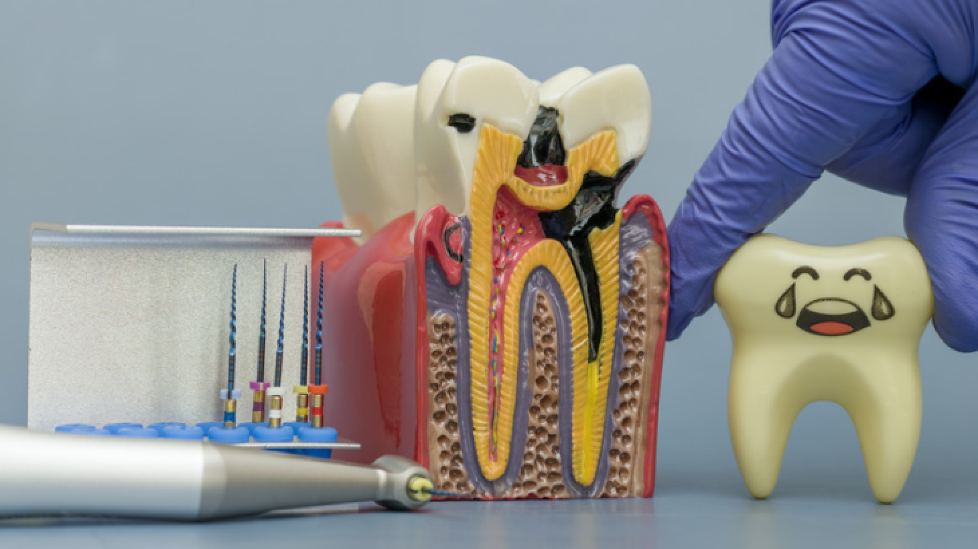
Side tooth decay, also known as interproximal tooth decay, occurs on the surfaces where two adjacent teeth touch. These areas are difficult for toothbrush bristles to reach and are almost impossible to inspect with the naked eye. The primary cause is often inadequate flossing, which allows plaque to accumulate between the teeth. Dental X-rays are the most reliable way to detect decay in these areas, and fillings can treat it before it reaches the dental pulp.
Most people check their teeth by smiling in front of a mirror and looking for dark spots on the tooth surfaces. The problem is that this method cannot reliably detect side tooth decay because these cavities hide between two closely spaced teeth, in areas that neither toothbrush bristles nor the naked eye can reach.
As a result, many people only discover that they have side tooth decay when their dentist takes X-rays and points out three or four dark spots, even though the outer surfaces of their teeth still look clean and white.
This article explains why the spaces between teeth are particularly vulnerable to decay, which warning signs you may notice before visiting a dentist, how side tooth decay is treated, and what you can do to prevent it effectively.
What Is Side Tooth Decay, and How Is It Different from Other Cavities?
Each tooth has several surfaces, including the chewing surface with its grooves and pits, the outer surface facing the cheek, the inner surface facing the tongue, and the interproximal surfaces that face neighboring teeth. Decay that develops on these interproximal surfaces is commonly referred to as side tooth decay.
There are three key differences between side tooth decay and cavities on the chewing surfaces.
First, it is difficult to see because the affected area lies beneath the contact point between two adjacent teeth.
Second, toothbrush bristles cannot effectively reach the area, regardless of how long or vigorously you brush. The bristles are simply too large to clean the narrow space properly.
Third, when one tooth develops decay on its side, the adjacent tooth may also be affected around the same time because both surfaces are exposed to plaque in the same area.
Together, these factors mean that side tooth decay is often detected later than cavities in other locations. By the time it is discovered, the damage may be deeper than expected.
However, the underlying cause is the same as that of other cavities: acids produced by bacteria as they break down sugars. To learn more about the underlying process, read our guide on what causes tooth decay.
Why Are the Spaces Between Teeth So Prone to Decay?
The spaces between teeth create an environment that encourages bacteria and plaque to accumulate.
Less Saliva Reaches These Areas
Saliva is one of the body's most important natural defenses against tooth decay. It helps wash away food particles, neutralize acids, and restore minerals to tooth enamel.
However, saliva has limited access to narrow spaces between closely positioned teeth. As a result, acids can remain in these areas longer than they do on more exposed tooth surfaces.
Food Particles Easily Become Trapped
Whenever you chew, small food particles can become lodged between your teeth. If they are not removed, they can contribute to plaque accumulation and provide a favorable environment for bacteria.
If you repeatedly notice food getting trapped in the same space after meals, it may indicate an existing dental problem that needs attention.
Toothbrush Bristles Cannot Reach These Surfaces Effectively
Even when using an expensive electric toothbrush or brushing for five minutes, brushing alone cannot clean every tooth surface effectively. The surfaces between teeth require additional cleaning with dental floss or an appropriately sized interdental brush.
Crowded or Overlapping Teeth Increase the Risk
Crowded or overlapping teeth create narrow, irregular spaces that are more difficult to clean than normally aligned teeth. People with these conditions may therefore face a higher risk of side tooth decay.
The same applies to people whose teeth have tilted after losing a neighboring tooth without replacing it. These changes can create unusual spaces where food and plaque accumulate easily.
Learn more about the causes and treatment options for tilted teeth, a condition that can also make cleaning between teeth more difficult.
Warning Signs of Side Tooth Decay
Although side tooth decay is difficult to see, your mouth may provide indirect warning signs. If you notice any of the following symptoms, consider scheduling a dental examination and asking whether X-rays are appropriate.
Food repeatedly gets stuck in the same space. You may need to use dental floss or a toothpick to remove food after almost every meal.
Dental floss repeatedly frays or breaks in one particular space. This may indicate an irregular tooth surface, a rough restoration, or another issue that requires examination.
A particular space produces an unusually strong odor. If the odor persists when you smell the floss after cleaning that area, it may indicate trapped debris or another dental problem.
Your teeth feel sensitive to cold drinks or sweet foods. You may find it difficult to identify exactly which tooth is causing the sensitivity.
You notice a faint dark shadow beneath the enamel near the edge of a tooth. This may indicate changes beneath the enamel, although an examination is needed to determine the cause.
The gum between two teeth becomes red, swollen, or bleeds when you floss. This may be related to plaque buildup, gum inflammation, or another local problem.
The space between two teeth seems wider than before. Changes in tooth position or gum health may explain this, so it is worth having the area checked.
One important point is that the absence of symptoms does not mean you are free from side tooth decay. Early- and intermediate-stage cavities between teeth can develop without noticeable discomfort.
Bitewing dental X-rays are commonly used to detect decay between the upper and lower teeth. Your dentist will determine whether these images are necessary based on your dental history, symptoms, and risk of developing cavities.
Comparing Different Types of Tooth Decay
Understanding how cavities differ by location helps explain why dentists place particular importance on examining the spaces between teeth.
Location
Visible to the Naked Eye
Accessible to a Toothbrush
Common Detection Stage
Filling Difficulty
Chewing surface
Often visible
Generally accessible
Early to intermediate
Usually straightforward
Side surface or between teeth
Often difficult to see
Difficult to reach effectively
May be detected at an intermediate or advanced stage
More challenging; requires careful access and contact-point reconstruction
Near the gumline
May be visible when the lip is lifted
Accessible with proper brushing
Often detected early
Moderate; moisture control is important
Beneath an existing filling
Usually not directly visible
Not applicable
May be detected after further damage develops
More complex; the existing restoration may need to be removed
As the table shows, side tooth decay presents several challenges: it is difficult to see, difficult to clean, and may not be detected until it has progressed. This is why flossing is not merely an optional addition to oral hygiene; it is an essential part of cleaning between teeth.
How Do Dentists Detect Side Tooth Decay?
Because visual inspection and probing alone may not be sufficient to identify decay between teeth, dentists use a combination of examination methods.
One commonly used method is a bitewing X-ray. During this procedure, you bite down on a small image receptor positioned between your upper and lower teeth. The resulting image shows the crowns of the teeth and the areas between them.
Decay may appear as a dark, often triangular-shaped area extending inward from the side of a tooth. Your dentist will assess whether the lesion is limited to the enamel, has reached the dentin, or is approaching the dental pulp. This information helps determine whether treatment is needed immediately or whether monitoring may be appropriate.
For front teeth, dentists may also use light-based examination techniques to identify areas that appear darker beneath the tooth surface. Dental floss can provide additional information if it repeatedly catches, frays, or breaks in a particular area.
These methods can support the examination, but X-rays are often particularly useful for detecting cavities that are hidden between adjacent teeth. No single method is perfect, so your dentist will interpret the findings alongside your symptoms and clinical examination.
How Is Side Tooth Decay Treated According to Its Severity?
Treatment depends on how deeply the decay has progressed, not simply on whether you have symptoms.
Stage 1: Decay Is Limited to the Enamel
If the affected area remains within the enamel, your dentist may recommend monitoring rather than drilling immediately, depending on the appearance and activity of the lesion.
Treatment may include professional fluoride application, improved daily cleaning between teeth, and dietary changes to reduce frequent sugar exposure. Your dentist may arrange follow-up examinations or X-rays when clinically appropriate to determine whether the lesion has remained stable or progressed.
This approach aims to preserve as much healthy tooth structure as possible. However, not every enamel lesion can be reversed, so the decision should be based on a professional assessment.
Stage 2: Decay Has Reached the Dentin
Once decay reaches the dentin, it may progress more readily because dentin is less resistant to acid than enamel. A filling is often necessary at this stage.
Restoring a cavity between teeth requires careful work. The dentist must access the affected side without damaging the neighboring tooth. A wedge and a thin matrix band may be placed between the teeth to create a temporary wall before the filling material is applied and shaped.
The dentist must also recreate the correct contact point between the two teeth. If the contact is too loose, food may become trapped. If it is too tight, dental floss may be difficult to pass through.
For these reasons, filling a cavity between teeth can be more technically demanding than filling one on a chewing surface. If you are concerned about the procedure, read our guide to dental fillings.
Stage 3: Decay Has Reached the Dental Pulp
If the decay extends into the dental pulp, symptoms may include spontaneous throbbing pain, pain that worsens at night, or prolonged sensitivity. However, symptoms vary, and their absence does not rule out significant damage.
At this stage, a simple filling may no longer be sufficient. Root canal treatment may be needed to remove infected or irreversibly inflamed pulp tissue, followed by restoration of the tooth. Depending on how much tooth structure remains, a crown may also be recommended.
If the decay extends far below the gumline and the tooth cannot be predictably restored, extraction may be considered. Your dentist can then discuss suitable replacement options.
Read more about tooth extraction and the factors that influence this decision.
Pros and Cons of Filling a Cavity Between Teeth
Understanding the benefits and limitations of treatment helps you set realistic expectations and care for your teeth after a filling.
The main benefit is that removing the decayed tissue and restoring the tooth can stop the existing cavity from progressing. A properly shaped restoration can also restore the contact between adjacent teeth and make the area easier to maintain. When decay is treated early, a filling is generally less complex and less expensive than root canal treatment.
However, the spaces between teeth remain challenging areas to clean, so recurrent decay can develop around the edges of a restoration if plaque continues to accumulate.
Some people experience temporary sensitivity for a short period after a filling. The contact between the restored tooth and its neighbor may also need adjustment if floss feels excessively tight or passes through too easily.
These issues should not be ignored. Contact your dentist if sensitivity persists, your bite feels uneven, or food continues to get trapped in the same space after treatment.
How to Prevent Side Tooth Decay Effectively
Because toothbrush bristles cannot adequately clean the surfaces between teeth, prevention requires tools specifically designed for these areas. Follow these steps every day, particularly before bedtime.
Cut approximately 45 cm of dental floss. Wrap the ends around your middle fingers, leaving about 2–3 cm of floss available for cleaning.
Gently guide the floss between your teeth. Use a controlled back-and-forth motion rather than snapping it down, which can injure your gums.
Curve the floss into a C shape around one tooth. Move it gently up and down along the tooth surface, extending slightly beneath the gumline without forcing it.
Clean the neighboring tooth in the same space. Each gap has two tooth surfaces that need to be cleaned.
Move to a clean section of floss for the next gap. This helps avoid transferring debris from one area to another.
Clean every space, including behind the last molars. These areas are often overlooked.
Use an interdental brush when appropriate. People with wider spaces, receding gums, or orthodontic appliances may benefit from an appropriately sized interdental brush in addition to flossing.
Brush with fluoride toothpaste afterward. Spit out the excess toothpaste and avoid repeatedly rinsing with water so that more fluoride remains on the tooth surfaces.
You should also reduce how frequently you consume sugary drinks and snacks throughout the day. Frequent sugar exposure encourages acid production, increasing the risk of decay.
The timing of dental X-rays should be individualized according to your risk level. Your dentist can recommend an appropriate interval based on your cavity history, clinical findings, and previous X-ray results.
Who Should Pay Special Attention to Side Tooth Decay?
The following groups may benefit from paying particular attention to cleaning between teeth and attending regular dental examinations.
People who rarely or never floss
If you have never used dental floss or use it inconsistently, plaque may have accumulated between your teeth without your knowledge. Arrange a dental examination and ask whether bitewing X-rays are appropriate. Start cleaning between your teeth daily.
People with crowded or overlapping teeth
Crowded teeth create spaces that can be difficult to clean. If you have lost a tooth and have not replaced it, changes in the position of neighboring teeth may also make cleaning more challenging. An interdental brush may help when appropriately sized, and your dentist may recommend more frequent monitoring.
People undergoing orthodontic treatment or who have recently had braces removed
Braces and other orthodontic appliances make effective cleaning more difficult. If plaque accumulates around the teeth and between adjacent surfaces, cavities may develop during treatment and become more apparent after the appliances are removed.
Consistent oral hygiene and regular dental examinations can help protect the time and money invested in orthodontic treatment.
Conclusion
Side tooth decay develops on the surfaces where two adjacent teeth touch. These areas are difficult for toothbrush bristles to reach, receive less direct saliva flow, and are often impossible to inspect with the naked eye. As a result, cavities in these locations may be detected later and be more advanced than expected.
Warning signs can include food repeatedly getting trapped in the same space, dental floss fraying or breaking in one particular area, or an unusual localized odor. However, cavities can develop without noticeable symptoms, so a dental examination and appropriate X-rays may be necessary to identify them.
Treatment depends on the depth of the decay. Early enamel lesions may be monitored and managed with preventive measures, cavities that reach the dentin often require fillings, and damage involving the dental pulp may require root canal treatment or, in some cases, extraction.
The most important preventive step is to clean between your teeth properly every day using dental floss or an appropriately sized interdental brush.
If you have not had your teeth examined or had your risk of decay assessed for several years, or if food repeatedly becomes trapped in the same space, a dental examination can provide clearer answers than looking in the mirror alone.
At Pasook Dental Clinic, digital dental X-ray equipment can help identify cavities between teeth, including lesions that may be difficult to see during a visual examination. The dental team can explain the findings and discuss suitable treatment options, including fillings that require careful reconstruction of the contact points between adjacent teeth. The clinic also offers clean, professional facilities across multiple branches for convenient access to dental care.
Frequently Asked Questions (FAQ)
1. Can I see side tooth decay myself by looking carefully in a mirror?
In some front teeth, you may notice a faint dark shadow beneath the enamel. However, cavities between molars—the teeth where chewing-surface and interproximal problems commonly occur—are often impossible to see directly because the affected area is hidden by the neighboring tooth. A dental examination and appropriate X-rays are often needed to confirm the diagnosis.
2. Should I stop flossing if my gums bleed?
Not necessarily. Bleeding may indicate gum inflammation caused by plaque buildup. If you floss gently and correctly every day, mild bleeding may improve within one to two weeks. However, if bleeding continues, is heavy, or is accompanied by pain or swelling, consult your dentist to identify the cause.
3. Why does food still get stuck between my teeth after a filling?
The contact point between the restored tooth and its neighbor may be too loose, allowing food to become trapped. This is not something you should simply tolerate. Tell your dentist so the restoration can be evaluated and adjusted or replaced if necessary. Leaving the problem unresolved may contribute to ongoing gum irritation and increase the difficulty of cleaning the area.
4. Does side tooth decay spread faster than cavities in other locations?
Not necessarily. However, cavities between teeth can go unnoticed for longer because they are difficult to see and clean. By the time they are detected, they may have progressed further or affected both neighboring teeth.
5. Can mouthwash replace dental floss?
No. Mouthwash can help freshen breath and, depending on its ingredients, provide additional benefits for oral health. However, it cannot reliably remove plaque that adheres to the surfaces between teeth. Dental floss or an appropriately sized interdental brush provides the physical cleaning needed to remove plaque from these areas.
6. How often should I have X-rays to check for cavities between my teeth?
The appropriate interval depends on your individual risk. Adults with a low risk of tooth decay may need X-rays less frequently, while people with new or recurring cavities, crowded teeth, dry mouth, or orthodontic appliances may need more frequent examinations and imaging.
Your dentist will recommend an interval based on your dental history, current oral health, and previous X-ray findings rather than following the same schedule for everyone.
Most people check their teeth by smiling in front of a mirror and looking for dark spots on the tooth surfaces. The problem is that this method cannot reliably detect side tooth decay because these cavities hide between two closely spaced teeth, in areas that neither toothbrush bristles nor the naked eye can reach.
As a result, many people only discover that they have side tooth decay when their dentist takes X-rays and points out three or four dark spots, even though the outer surfaces of their teeth still look clean and white.
This article explains why the spaces between teeth are particularly vulnerable to decay, which warning signs you may notice before visiting a dentist, how side tooth decay is treated, and what you can do to prevent it effectively.
What Is Side Tooth Decay, and How Is It Different from Other Cavities?
Each tooth has several surfaces, including the chewing surface with its grooves and pits, the outer surface facing the cheek, the inner surface facing the tongue, and the interproximal surfaces that face neighboring teeth. Decay that develops on these interproximal surfaces is commonly referred to as side tooth decay.
There are three key differences between side tooth decay and cavities on the chewing surfaces.
First, it is difficult to see because the affected area lies beneath the contact point between two adjacent teeth.
Second, toothbrush bristles cannot effectively reach the area, regardless of how long or vigorously you brush. The bristles are simply too large to clean the narrow space properly.
Third, when one tooth develops decay on its side, the adjacent tooth may also be affected around the same time because both surfaces are exposed to plaque in the same area.
Together, these factors mean that side tooth decay is often detected later than cavities in other locations. By the time it is discovered, the damage may be deeper than expected.
However, the underlying cause is the same as that of other cavities: acids produced by bacteria as they break down sugars. To learn more about the underlying process, read our guide on what causes tooth decay.
Why Are the Spaces Between Teeth So Prone to Decay?
The spaces between teeth create an environment that encourages bacteria and plaque to accumulate.
Less Saliva Reaches These Areas
Saliva is one of the body's most important natural defenses against tooth decay. It helps wash away food particles, neutralize acids, and restore minerals to tooth enamel.
However, saliva has limited access to narrow spaces between closely positioned teeth. As a result, acids can remain in these areas longer than they do on more exposed tooth surfaces.
Food Particles Easily Become Trapped
Whenever you chew, small food particles can become lodged between your teeth. If they are not removed, they can contribute to plaque accumulation and provide a favorable environment for bacteria.
If you repeatedly notice food getting trapped in the same space after meals, it may indicate an existing dental problem that needs attention.
Toothbrush Bristles Cannot Reach These Surfaces Effectively
Even when using an expensive electric toothbrush or brushing for five minutes, brushing alone cannot clean every tooth surface effectively. The surfaces between teeth require additional cleaning with dental floss or an appropriately sized interdental brush.
Crowded or Overlapping Teeth Increase the Risk
Crowded or overlapping teeth create narrow, irregular spaces that are more difficult to clean than normally aligned teeth. People with these conditions may therefore face a higher risk of side tooth decay.
The same applies to people whose teeth have tilted after losing a neighboring tooth without replacing it. These changes can create unusual spaces where food and plaque accumulate easily.
Learn more about the causes and treatment options for tilted teeth, a condition that can also make cleaning between teeth more difficult.
Warning Signs of Side Tooth Decay
Although side tooth decay is difficult to see, your mouth may provide indirect warning signs. If you notice any of the following symptoms, consider scheduling a dental examination and asking whether X-rays are appropriate.
Food repeatedly gets stuck in the same space. You may need to use dental floss or a toothpick to remove food after almost every meal.
Dental floss repeatedly frays or breaks in one particular space. This may indicate an irregular tooth surface, a rough restoration, or another issue that requires examination.
A particular space produces an unusually strong odor. If the odor persists when you smell the floss after cleaning that area, it may indicate trapped debris or another dental problem.
Your teeth feel sensitive to cold drinks or sweet foods. You may find it difficult to identify exactly which tooth is causing the sensitivity.
You notice a faint dark shadow beneath the enamel near the edge of a tooth. This may indicate changes beneath the enamel, although an examination is needed to determine the cause.
The gum between two teeth becomes red, swollen, or bleeds when you floss. This may be related to plaque buildup, gum inflammation, or another local problem.
The space between two teeth seems wider than before. Changes in tooth position or gum health may explain this, so it is worth having the area checked.
One important point is that the absence of symptoms does not mean you are free from side tooth decay. Early- and intermediate-stage cavities between teeth can develop without noticeable discomfort.
Bitewing dental X-rays are commonly used to detect decay between the upper and lower teeth. Your dentist will determine whether these images are necessary based on your dental history, symptoms, and risk of developing cavities.
Comparing Different Types of Tooth Decay
Understanding how cavities differ by location helps explain why dentists place particular importance on examining the spaces between teeth.
Location
Visible to the Naked Eye
Accessible to a Toothbrush
Common Detection Stage
Filling Difficulty
Chewing surface
Often visible
Generally accessible
Early to intermediate
Usually straightforward
Side surface or between teeth
Often difficult to see
Difficult to reach effectively
May be detected at an intermediate or advanced stage
More challenging; requires careful access and contact-point reconstruction
Near the gumline
May be visible when the lip is lifted
Accessible with proper brushing
Often detected early
Moderate; moisture control is important
Beneath an existing filling
Usually not directly visible
Not applicable
May be detected after further damage develops
More complex; the existing restoration may need to be removed
As the table shows, side tooth decay presents several challenges: it is difficult to see, difficult to clean, and may not be detected until it has progressed. This is why flossing is not merely an optional addition to oral hygiene; it is an essential part of cleaning between teeth.
How Do Dentists Detect Side Tooth Decay?
Because visual inspection and probing alone may not be sufficient to identify decay between teeth, dentists use a combination of examination methods.
One commonly used method is a bitewing X-ray. During this procedure, you bite down on a small image receptor positioned between your upper and lower teeth. The resulting image shows the crowns of the teeth and the areas between them.
Decay may appear as a dark, often triangular-shaped area extending inward from the side of a tooth. Your dentist will assess whether the lesion is limited to the enamel, has reached the dentin, or is approaching the dental pulp. This information helps determine whether treatment is needed immediately or whether monitoring may be appropriate.
For front teeth, dentists may also use light-based examination techniques to identify areas that appear darker beneath the tooth surface. Dental floss can provide additional information if it repeatedly catches, frays, or breaks in a particular area.
These methods can support the examination, but X-rays are often particularly useful for detecting cavities that are hidden between adjacent teeth. No single method is perfect, so your dentist will interpret the findings alongside your symptoms and clinical examination.
How Is Side Tooth Decay Treated According to Its Severity?
Treatment depends on how deeply the decay has progressed, not simply on whether you have symptoms.
Stage 1: Decay Is Limited to the Enamel
If the affected area remains within the enamel, your dentist may recommend monitoring rather than drilling immediately, depending on the appearance and activity of the lesion.
Treatment may include professional fluoride application, improved daily cleaning between teeth, and dietary changes to reduce frequent sugar exposure. Your dentist may arrange follow-up examinations or X-rays when clinically appropriate to determine whether the lesion has remained stable or progressed.
This approach aims to preserve as much healthy tooth structure as possible. However, not every enamel lesion can be reversed, so the decision should be based on a professional assessment.
Stage 2: Decay Has Reached the Dentin
Once decay reaches the dentin, it may progress more readily because dentin is less resistant to acid than enamel. A filling is often necessary at this stage.
Restoring a cavity between teeth requires careful work. The dentist must access the affected side without damaging the neighboring tooth. A wedge and a thin matrix band may be placed between the teeth to create a temporary wall before the filling material is applied and shaped.
The dentist must also recreate the correct contact point between the two teeth. If the contact is too loose, food may become trapped. If it is too tight, dental floss may be difficult to pass through.
For these reasons, filling a cavity between teeth can be more technically demanding than filling one on a chewing surface. If you are concerned about the procedure, read our guide to dental fillings.
Stage 3: Decay Has Reached the Dental Pulp
If the decay extends into the dental pulp, symptoms may include spontaneous throbbing pain, pain that worsens at night, or prolonged sensitivity. However, symptoms vary, and their absence does not rule out significant damage.
At this stage, a simple filling may no longer be sufficient. Root canal treatment may be needed to remove infected or irreversibly inflamed pulp tissue, followed by restoration of the tooth. Depending on how much tooth structure remains, a crown may also be recommended.
If the decay extends far below the gumline and the tooth cannot be predictably restored, extraction may be considered. Your dentist can then discuss suitable replacement options.
Read more about tooth extraction and the factors that influence this decision.
Pros and Cons of Filling a Cavity Between Teeth
Understanding the benefits and limitations of treatment helps you set realistic expectations and care for your teeth after a filling.
The main benefit is that removing the decayed tissue and restoring the tooth can stop the existing cavity from progressing. A properly shaped restoration can also restore the contact between adjacent teeth and make the area easier to maintain. When decay is treated early, a filling is generally less complex and less expensive than root canal treatment.
However, the spaces between teeth remain challenging areas to clean, so recurrent decay can develop around the edges of a restoration if plaque continues to accumulate.
Some people experience temporary sensitivity for a short period after a filling. The contact between the restored tooth and its neighbor may also need adjustment if floss feels excessively tight or passes through too easily.
These issues should not be ignored. Contact your dentist if sensitivity persists, your bite feels uneven, or food continues to get trapped in the same space after treatment.
How to Prevent Side Tooth Decay Effectively
Because toothbrush bristles cannot adequately clean the surfaces between teeth, prevention requires tools specifically designed for these areas. Follow these steps every day, particularly before bedtime.
Cut approximately 45 cm of dental floss. Wrap the ends around your middle fingers, leaving about 2–3 cm of floss available for cleaning.
Gently guide the floss between your teeth. Use a controlled back-and-forth motion rather than snapping it down, which can injure your gums.
Curve the floss into a C shape around one tooth. Move it gently up and down along the tooth surface, extending slightly beneath the gumline without forcing it.
Clean the neighboring tooth in the same space. Each gap has two tooth surfaces that need to be cleaned.
Move to a clean section of floss for the next gap. This helps avoid transferring debris from one area to another.
Clean every space, including behind the last molars. These areas are often overlooked.
Use an interdental brush when appropriate. People with wider spaces, receding gums, or orthodontic appliances may benefit from an appropriately sized interdental brush in addition to flossing.
Brush with fluoride toothpaste afterward. Spit out the excess toothpaste and avoid repeatedly rinsing with water so that more fluoride remains on the tooth surfaces.
You should also reduce how frequently you consume sugary drinks and snacks throughout the day. Frequent sugar exposure encourages acid production, increasing the risk of decay.
The timing of dental X-rays should be individualized according to your risk level. Your dentist can recommend an appropriate interval based on your cavity history, clinical findings, and previous X-ray results.
Who Should Pay Special Attention to Side Tooth Decay?
The following groups may benefit from paying particular attention to cleaning between teeth and attending regular dental examinations.
People who rarely or never floss
If you have never used dental floss or use it inconsistently, plaque may have accumulated between your teeth without your knowledge. Arrange a dental examination and ask whether bitewing X-rays are appropriate. Start cleaning between your teeth daily.
People with crowded or overlapping teeth
Crowded teeth create spaces that can be difficult to clean. If you have lost a tooth and have not replaced it, changes in the position of neighboring teeth may also make cleaning more challenging. An interdental brush may help when appropriately sized, and your dentist may recommend more frequent monitoring.
People undergoing orthodontic treatment or who have recently had braces removed
Braces and other orthodontic appliances make effective cleaning more difficult. If plaque accumulates around the teeth and between adjacent surfaces, cavities may develop during treatment and become more apparent after the appliances are removed.
Consistent oral hygiene and regular dental examinations can help protect the time and money invested in orthodontic treatment.
Conclusion
Side tooth decay develops on the surfaces where two adjacent teeth touch. These areas are difficult for toothbrush bristles to reach, receive less direct saliva flow, and are often impossible to inspect with the naked eye. As a result, cavities in these locations may be detected later and be more advanced than expected.
Warning signs can include food repeatedly getting trapped in the same space, dental floss fraying or breaking in one particular area, or an unusual localized odor. However, cavities can develop without noticeable symptoms, so a dental examination and appropriate X-rays may be necessary to identify them.
Treatment depends on the depth of the decay. Early enamel lesions may be monitored and managed with preventive measures, cavities that reach the dentin often require fillings, and damage involving the dental pulp may require root canal treatment or, in some cases, extraction.
The most important preventive step is to clean between your teeth properly every day using dental floss or an appropriately sized interdental brush.
If you have not had your teeth examined or had your risk of decay assessed for several years, or if food repeatedly becomes trapped in the same space, a dental examination can provide clearer answers than looking in the mirror alone.
At Pasook Dental Clinic, digital dental X-ray equipment can help identify cavities between teeth, including lesions that may be difficult to see during a visual examination. The dental team can explain the findings and discuss suitable treatment options, including fillings that require careful reconstruction of the contact points between adjacent teeth. The clinic also offers clean, professional facilities across multiple branches for convenient access to dental care.
Frequently Asked Questions (FAQ)
1. Can I see side tooth decay myself by looking carefully in a mirror?
In some front teeth, you may notice a faint dark shadow beneath the enamel. However, cavities between molars—the teeth where chewing-surface and interproximal problems commonly occur—are often impossible to see directly because the affected area is hidden by the neighboring tooth. A dental examination and appropriate X-rays are often needed to confirm the diagnosis.
2. Should I stop flossing if my gums bleed?
Not necessarily. Bleeding may indicate gum inflammation caused by plaque buildup. If you floss gently and correctly every day, mild bleeding may improve within one to two weeks. However, if bleeding continues, is heavy, or is accompanied by pain or swelling, consult your dentist to identify the cause.
3. Why does food still get stuck between my teeth after a filling?
The contact point between the restored tooth and its neighbor may be too loose, allowing food to become trapped. This is not something you should simply tolerate. Tell your dentist so the restoration can be evaluated and adjusted or replaced if necessary. Leaving the problem unresolved may contribute to ongoing gum irritation and increase the difficulty of cleaning the area.
4. Does side tooth decay spread faster than cavities in other locations?
Not necessarily. However, cavities between teeth can go unnoticed for longer because they are difficult to see and clean. By the time they are detected, they may have progressed further or affected both neighboring teeth.
5. Can mouthwash replace dental floss?
No. Mouthwash can help freshen breath and, depending on its ingredients, provide additional benefits for oral health. However, it cannot reliably remove plaque that adheres to the surfaces between teeth. Dental floss or an appropriately sized interdental brush provides the physical cleaning needed to remove plaque from these areas.
6. How often should I have X-rays to check for cavities between my teeth?
The appropriate interval depends on your individual risk. Adults with a low risk of tooth decay may need X-rays less frequently, while people with new or recurring cavities, crowded teeth, dry mouth, or orthodontic appliances may need more frequent examinations and imaging.
Your dentist will recommend an interval based on your dental history, current oral health, and previous X-ray findings rather than following the same schedule for everyone.
Related Content

Taking care of your teeth to prevent tooth decay is essential for your overall health. This article guides you through proper dental care and effective treatment to stop decay from progressing and avoid tooth loss.
What are the different types of wisdom teeth? Learn about tilted, impacted, partially erupted, and horizontal wisdom teeth, plus how to check which types are more likely to require surgical removal and which may not.

Wondering which wisdom teeth do not need surgery? Learn the characteristics of wisdom teeth that can be safely retained without removal, plus important warning signs and dental considerations.